36 Label Parts Of The Microscope Labels 2021

🎉 Main components of a light microscope. Parts of a microscope with
Parts of the Microscope (Labeled Diagrams) By Editorial Board December 14, 2022 The microscope is one of the must-have laboratory tools because of its ability to observe minute objects, usually living organisms that cannot be seen by the naked eyes. It is categorized into two: simple and compound microscopes.
Guide to understand microscope parts, names, functions & diagram
Base Microscope Worksheet The Light Microscope Light microscopes are used to examine cells at relatively low magnifications. Magnifications of about 2000X are the upper limit for light microscopes. The highest resolution of a light microscope is about 0.2 μm. The use of blue light to illuminate a specimen gives the highest resolution.
Light Microscope Main Parts Of Light Microscope Biology —
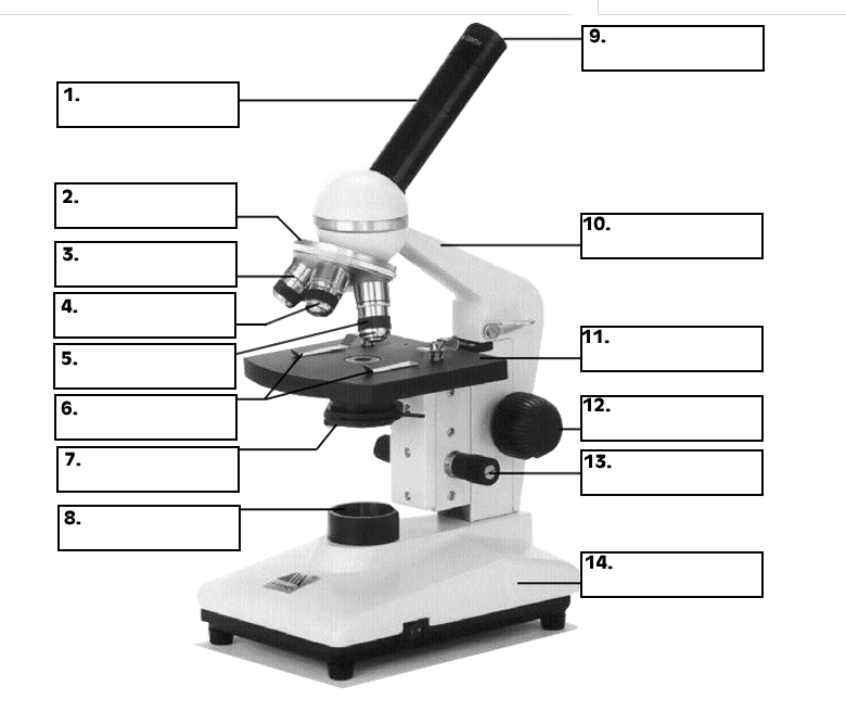
ACTIVITY Microscope parts In this activity, students identify and label the main parts of a microscope and describe their function. By the end of this activity, students should be able to:. READ MORE MORE Use this interactive to identify and label the main parts of a microscope. Drag and drop the text labels onto the microscope diagram.
Microscopes 7th Grade Science
The microscope illustrated in Figure 5 below was manufactured by Hugh Powell and Peter Lealand around 1850. The tripod base provided a sturdy support for the microscope, which many people consider the most advanced of its period. Parts of a Powell and Leland Microscope Diagram

Microscope Diagram Labeled, Unlabeled and Blank Parts of a Microscope
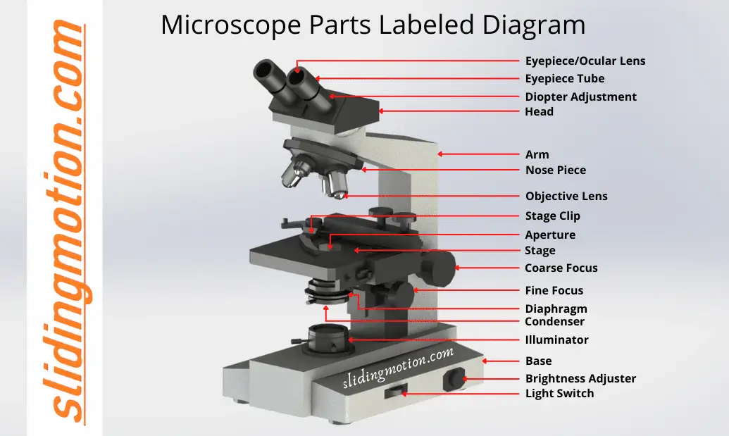
Parts of a Microscope and their Functions. The microscope comprises three main structural components: the head, the base, and the arm. HEAD: Also known as the body, the microscope's head houses the optical components in the upper portion. BASE- It serves as a support for microscopes. Illuminators for microscopic work are also included.

36 Label Parts Of The Microscope Labels 2021
The hand magnifying glass can magnify about 3 to 20×. Single-lensed simple microscopes can magnify up to 300×—and are capable of revealing bacteria —while compound microscopes can magnify up to 2,000×. A simple microscope can resolve below 1 micrometre (μm; one millionth of a metre); a compound microscope can resolve down to about 0.2 μm.

301 Moved Permanently
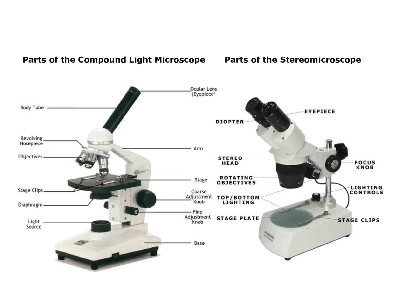
Parts of a Compound Microscope Each part of the compound microscope serves its own unique function, with each being important to the function of the scope as a whole. The individual parts of a compound microscope can vary heavily depending on the configuration & applications that the scope is being used for. Common compound microscope parts include: Compound Microscope Definitions for.

Diagrams of a Microscope 101 Diagrams
Iris diaphragm: Adjusts the amount of light that reaches the specimen. Condenser: Gathers and focuses light from the illuminator onto the specimen being viewed. Base: The base supports the microscope and it's where illuminator is located. How Does a Compound Microscope Work?
Microscope Parts Sketch at Explore collection of
2. Compound Microscope. Compound Microscope is a type of microscope that used visible light for illumination and multiple lenses system for magnification of specimen. Generally, it consists of two lenses; objective lens and ocular lens. It can magnify images up to 1000X.
Microscope Diagram to Print 101 Diagrams
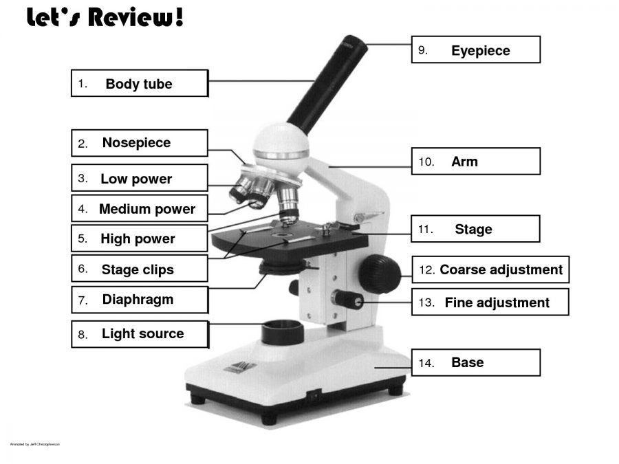
The main parts include the following: Fig: Parts of the microscope Labeled diagram Table of Contents Structural parts of a microscope: Optical parts of a microscope The Eyepiece The Eyepiece tube The objective lenses The focus knobs Stage The light source (Illuminator) Condenser The condenser focus knob The diaphragm

Microscope Diagram Labeled, Unlabeled and Blank Parts of a Microscope
To better understand the structure and function of a microscope, we need to take a look at the labeled microscope diagrams of the compound and electron microscope. These diagrams clearly explain the functioning of the microscopes along with their respective parts. Man's curiosity has led to great inventions. The microscope is one of them.
Microscope Diagram to Print 101 Diagrams
1. Eyepiece Lens and Eyepiece Tube 2. Objective Lens 3. Tube 4. Base 5.
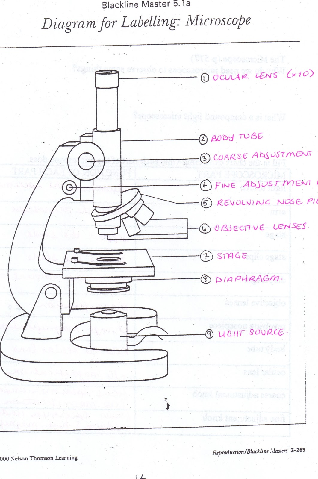
All Saints Online Diagram for Labelling Microscope
There are 1000 millimeters (mm) in one meter. 1 mm = 10 -3 meter. There are 1000 micrometers (microns, or µm) in one millimeter. 1 µm = 10 -6 meter. There are 1000 nanometers in one micrometer. 1 nm = 10 -9 meter. Figure 1: Resolving Power of Microscopes. The microscope is one of the microbiologist's greatest tools.

Parts of a microscope with functions and labeled diagram
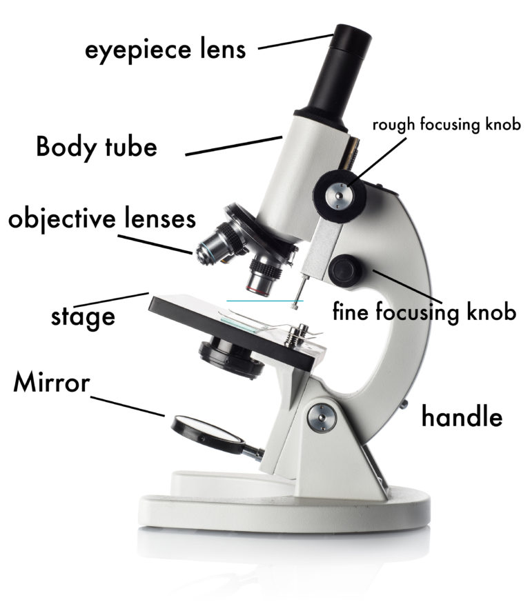
Tube: Connects the eyepiece to the objective lenses. Arm: Supports the tube and connects it to the base. Base: The bottom of the microscope, used for support. Illuminator: A steady light source (110 volts) used in place of a mirror. If your microscope has a mirror, it is used to reflect light from an external light source up through the bottom.

Diagrams of Microscope 101 Diagrams
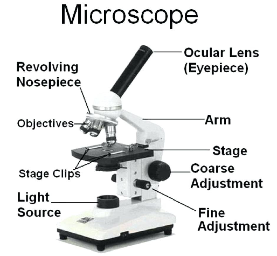
The 16 core parts of a compound microscope are: Head (Body) Arm Base Eyepiece Eyepiece tube Objective lenses Revolving Nosepiece (Turret) Rack stop Coarse adjustment knobs Fine adjustment knobs Stage Stage clips Aperture Illuminator Condenser Diaphragm Video: Parts of a compound Microscope with Diagram Explained
How to Use a Microscope
The field diaphragm control is located around the lens located in the base. Hinge Screw -This screw fixes the arm to the base and allow for the tilting of the arm. Stage Clips - They hold the slide firmly onto the stage. On/OFF Switch - This switch on the base of the microscope turns the illuminator off and on.
