Entamoeba histolytica Histopathology.guru
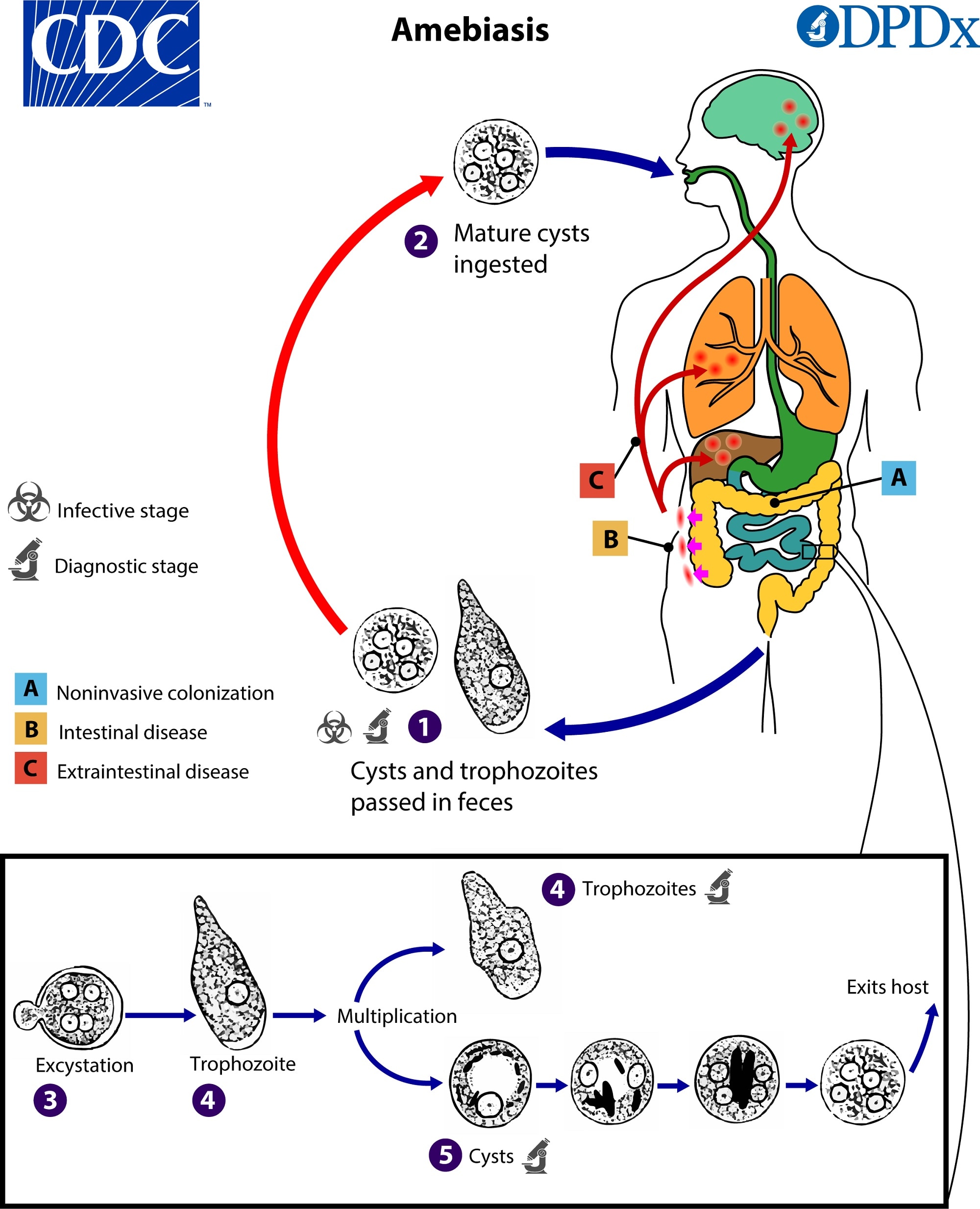
Life cycle of entamoeba histolytica diagram
Entamoeba histolytica is a pathogenic parasite in the intestine of human beings and many other primates. It inhabits the mucous and sub-mucous layers of the large intestine. It feeds mainly on the tissues of the intestinal wall and often produces severe ulcers and abscesses.

Public Domain Picture Entamoeba histolytica cyst. ID
Entamoeba histolytica is an anaerobic parasitic amoebozoan, part of the genus Entamoeba. [1] Predominantly infecting humans and other primates causing amoebiasis, E. histolytica is estimated to infect about 35-50 million people worldwide. [1] E. histolytica infection is estimated to kill more than 55,000 people each year. [2]

Life Cycle of Entamoeba histolytica. The human host ingests the
Entamoeba histolytica is well recognized as a pathogenic ameba, associated with intestinal and extraintestinal infections. Other morphologically-identical Entamoeba spp., including E. dispar, E. moshkovskii, and E. bangladeshi, are generally not associated with disease although investigations into pathogenic potential are ongoing.

Life cycle and pathogenicity of Entamoeba histolytica
Entamoeba histolytica is a protozoan that causes intestinal amebiasis as well as extraintestinal manifestations. Although 90 percent of E. histolytica infections are asymptomatic, nearly 50 million people become symptomatic, with about 100,000 deaths yearly. [1] Amebic infections are more prevalent in countries with lower socioeconomic conditions.

Traveling Small with a Nucleus Organism Entamoeba histolytica
Entamoeba histolytica, the etiological agent of amebiasis, is a major parasitic cause of morbidity and death, particularly in developing countries. It is estimated that around 50 million symptomatic cases and 100,000 deaths worldwide/year. [ 1]

Pin on Hand drawing
Entamoeba histolytica is an enteric protozoan parasite with worldwide distribution. It is responsible for amoebic dysentery (bloody diarrhea) and invasive extraintestinal amebiasis (such as liver abscess, peritonitis, and pleuropulmonary abscess).
Differences Between Entamoeba histolytica and Entamoeba coli
Protozoan species, genus Entamoeba. E. histolytica is morphologically similar to E. dispar, E. moshkovskii, and E. bangladeshi. Histolytic - histo + lyein (Greek) to loosen, trophozoites contain cytolytic and proteolytic enzymes, including collagenase and proteinases, and lyze neutrophils and macrophages. Cysts are ingested; excystation occurs.
ENTAMOEBA PARAZİTOLOJİ BİLGİPEDİA
Find Entamoeba Histolytica Drawing stock photos and editorial news pictures from Getty Images. Select from premium Entamoeba Histolytica Drawing of the highest quality.

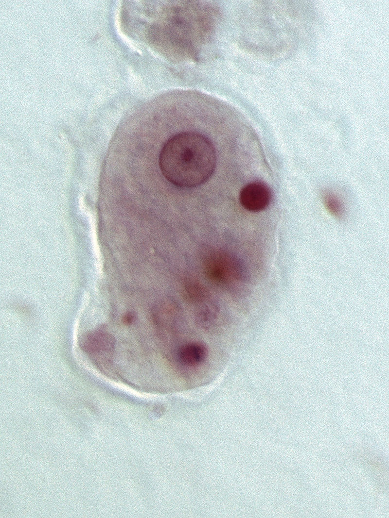
Entamoeba histolytica trophozoite and cyst microscopic view ((with
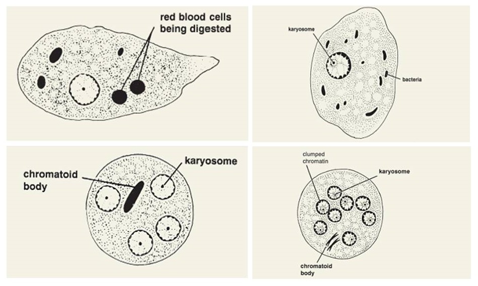
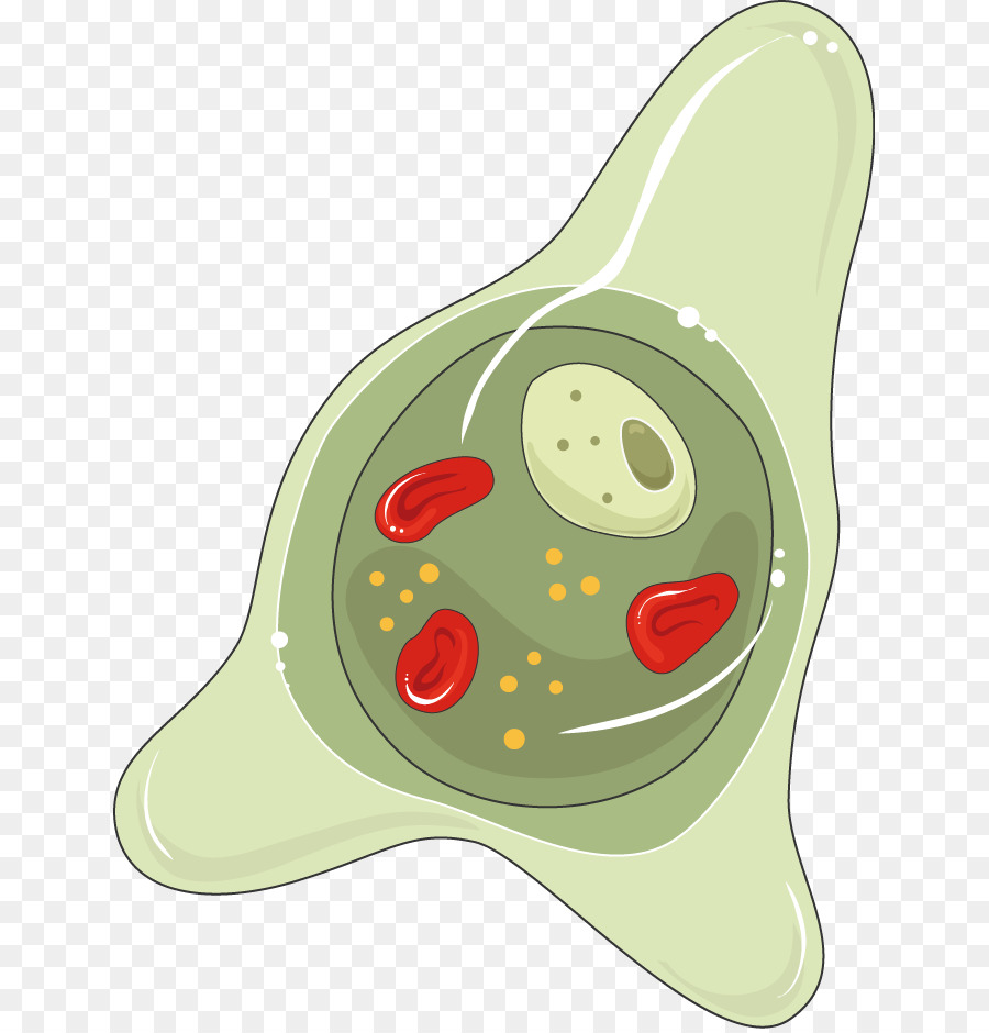
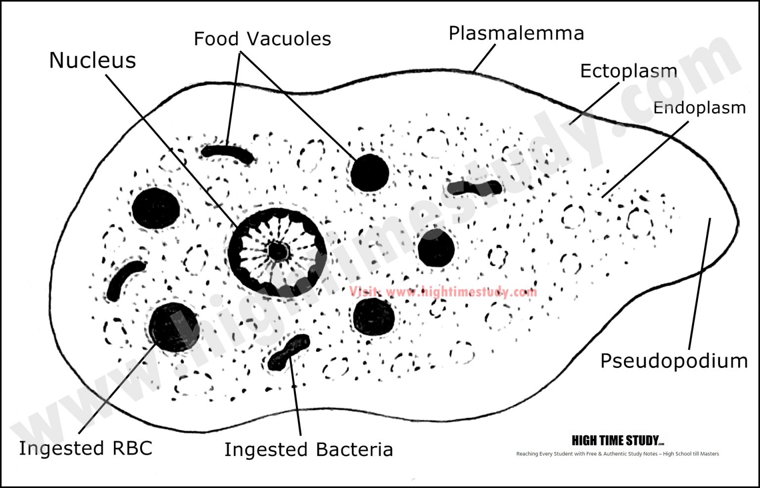
Structure of Entamoeba Histolytica: The amoeba has three stages in its life cycle, viz. the trophozoite stage, the precystic stage and the cystic stage. A. The trophozoite amoeba: ADVERTISEMENTS: This is the growing or feeding stage of the parasite having the following features: 1.
Entamoeba histolytica wikidoc
Entamoeba histolytica. Entamoeba histolytica is one of a number of species of small amoebae which live in the alimentary canal of humans. These are usually harmless protozoa, feeding on bacteria and particles in the intestine. In certain conditions, entamoeba invades the wall of the intestine or rectum causing ulceration and bleeding, with pain.

Entamoeba histolytica
Amebiasis is defined as infection with Entamoeba histolytica, regardless of associated symptomatology. In resource-rich nations, this parasitic protozoan is seen primarily in travelers to and emigrants from endemic areas. Infections range from asymptomatic colonization to amebic colitis and life-threatening abscesses. Importantly, disease may occur months to years after exposure. Although E.
A Entamoeba Histolytica, Parasitologia, Trophozoite png transparente grátis
how to draw entamoeba histolytica
Entamoeba histolytica Histopathology.guru
Browse Getty Images' premium collection of high-quality, authentic Entamoeba Histolytica Drawing stock photos, royalty-free images, and pictures. Entamoeba Histolytica Drawing stock photos are available in a variety of sizes and formats to fit your needs.
Entamoeba Histolytica Drawing
5 NEXT Browse Getty Images' premium collection of high-quality, authentic Entamoeba Histolytica Drawing stock photos, royalty-free images, and pictures. Entamoeba Histolytica Drawing stock photos are available in a variety of sizes and formats to fit your needs.

Figure 1 from ASPECTS ENTAMOEBA HISTOLYTICA Semantic Scholar
12.8K subscribers Subscribe 9.1K views 1 year ago This video will be very useful for students to draw the structure of entamoeba histolytica very easily. Thanks for watching and please.

15.19F Amoebic Dysentery (Amoebiasis) Biology LibreTexts
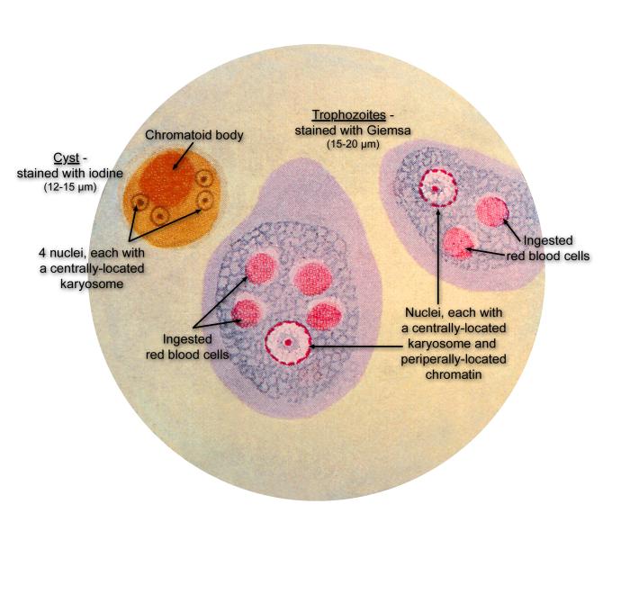
Wet mount. Entamoeba histolytica and Entamoeba dispar are morphologically identical species. In bright-field microscopy, E. histolytica/E. dispar cysts are spherical and usually measure 12 to 15 μm (range may be 10 to 20 μm). A mature cyst has 4 nuclei while an immature cyst may contain only 1 to 3 nuclei. Peripheral chromatin is fine.
