Flexor Digitorum Superficialis Anatomy Origin, Insertion, Action The Wellness Digest
Flexor Digitorum Superficialis Anatomy Origin, Insertion, Action The Wellness Digest
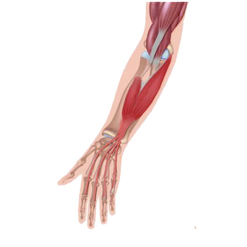
It is the bulk of muscle located at the superficial volar/anterior aspect of the forearm. [1] The flexor digitorum superficialis courses along the volar aspect of the forearm, superficial to the flexor digitorum profundus and flexor pollicis longus muscles, and deep to the palmaris longus, flexor carpi radialis, flexor carpi ulnaris, and.
Flexor Digitorum Superficialis Learn Muscles
The muscle-tendon arrangement of the m. flexor digitorum superficialis (FDS) varies among different primate groups. Recent developmental investigations revealed that the primordium of FDS emerges in the hand region first and relocates to the forearm later. The relationship between the diverse muscle-tendon arrangement and the characteristic.
Flexor Digitorum Superficialis Flexor Digitorum Superficialis to Anterior Interosseous
Flexor digitorum superficialis is innervated by the median nerve (C8-T1) and vascularized by the ulnar and radial arteries. The prime function of flexor digitorum superficialis is flexion of the digits 2-5 at the PIP and MCP joints. In addition, it contributes to the flexion of the hand at the wrist joint.

Anatomy Stock Images forearmhandmusculusflexordigitorumsuperficialismusclemiddle
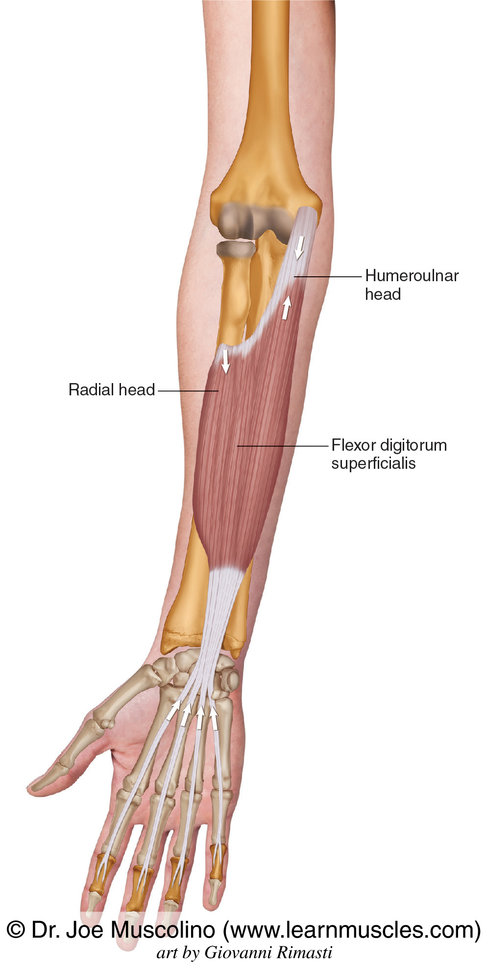
Flexor Digitorum Superficialis. Origin: Humeroulnar head: medial epicondyle of humerus, ulnar collateral ligament, and coronoid process of ulna; Radial head: superior half of anterior border of radius. Insertion: Bodies of middle phalanges of digits 2 - 5.

Flexor Digitorum Superficialis Muscle GetBodySmart
The m. flexor digitorum superficialis (FDS) in the common marmoset (right side of specimen no. 3). (a) Palmar view of the FDS and supplying nerves. Scale bar: 5 mm. Abbreviations of muscles allocated to nerve branches denote nerve branches to respective muscles. (b) Line drawing corresponding to the image in (a).
Flexor Digitorum Superficialis Muscle GetBodySmart
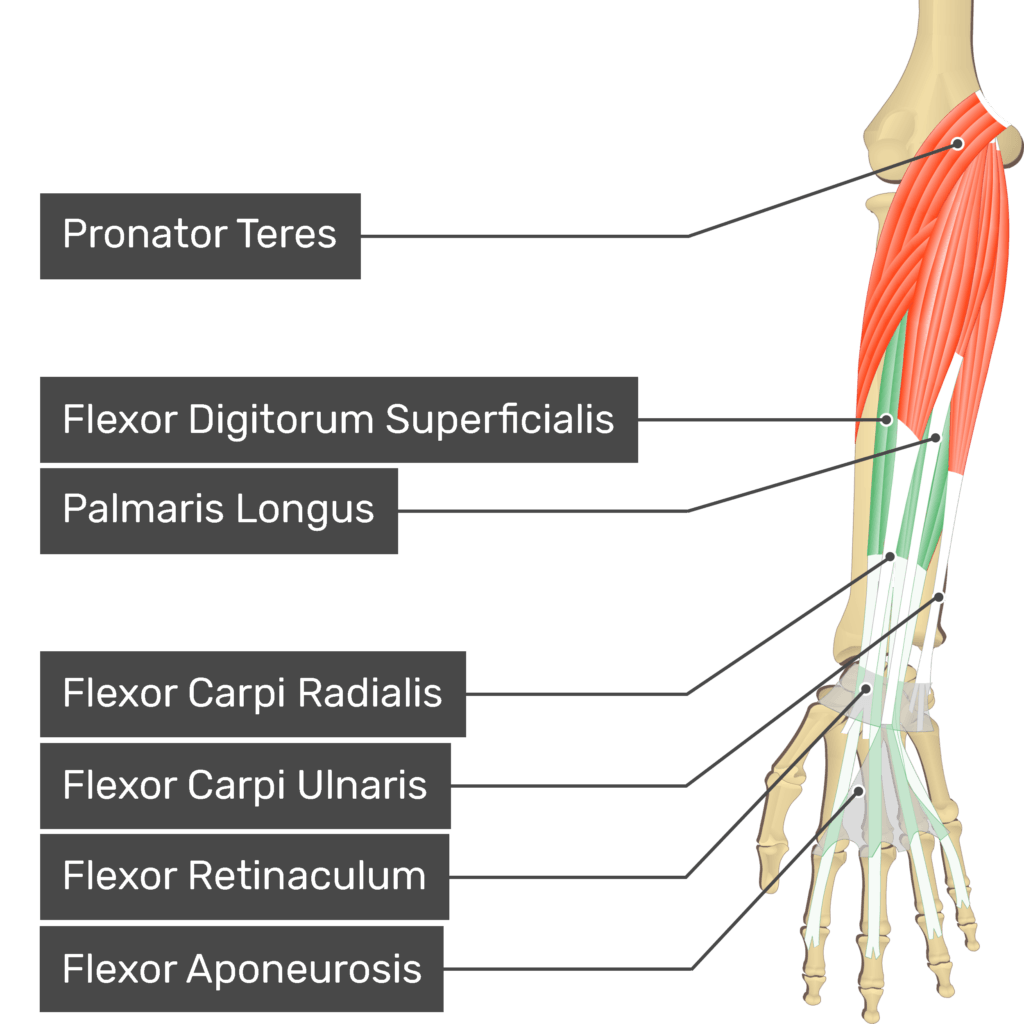
Flexor digitorum superficialis ( flexor digitorum sublimis) is an extrinsic flexor muscle of the fingers at the proximal interphalangeal joints . It is in the anterior compartment of the forearm. It is sometimes considered to be the deepest part of the superficial layer of this compartment, [1] [2] and sometimes considered to be a distinct.

FLEXOR DIGITORUM (SUPERFICIALIS) =deep to palmaris longus anterior of forearm ORIGIN medial
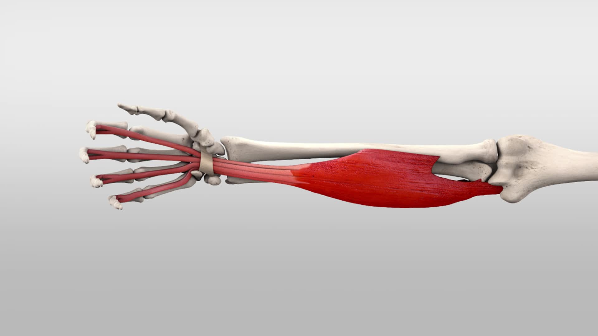
Flexor digitorum superficialis (FDS) muscle, also known as flexor digitorum sublimis muscle, is a muscle in the second (intermediate) layer of the anterior compartment of the forearm.It splits into four tendons, passes through the carpal tunnel under the flexor retinaculum.At the level of the head of the metacarpal, the flexor digitorum superficialis tendons split and wrap around the flexor.
Musculus flexor digitorum superficialis DocCheck
The flexor digitorum superficialis is an extrinsic muscle that allows the four medial fingers of the hand to flex. These fingers include the index, middle, ring, and pinkie fingers. The term.
Musculus flexor digitorum superficialis Anatomie Kenhub
The flexor digitorum superficialis and the flexor digitorium profundus tendons should be tested individually. A normal intact flexor digitorum superficialis is indicated when all the adjacent digits are held with all joints in extension while the patient flexes the finger at the proximal interphalangeal joint. On the other hand if the middle.
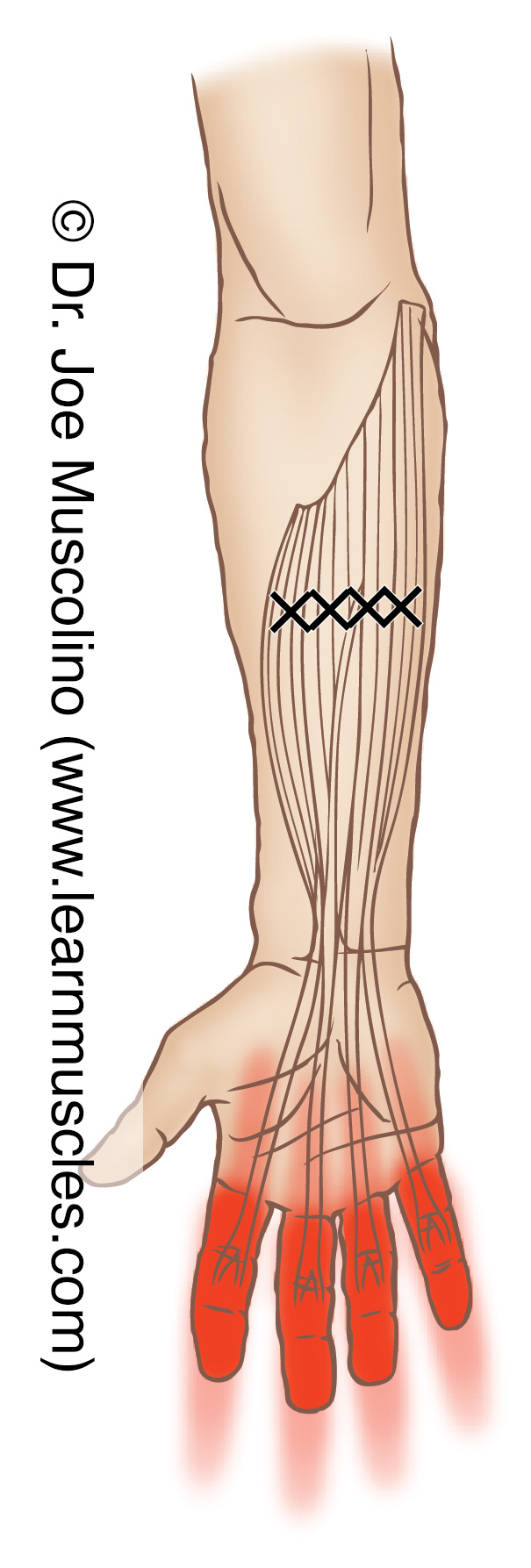
Flexors Digitorum Superficialis and Profundus Trigger Points Learn Muscles
The flexor digitorum superficialis (FDS), formerly known as the flexor digitorum sublimis, is the largest of the extrinsic flexors of the forearm. It forms the intermediate muscle layer between the superficial and deep muscle groups of the forearm.[1] It contains four digital components, with a tendon that inserts onto each corresponding finger. The flexor digitorum superficialis is the.

Repin to revise the muscle facts about the flexor digitorum superficialis muscle with Kenhub
1. Introduction. The flexor digitorum superficialis (FDS) is the largest among the superficial flexor muscles situated in the anterior compartment of the forearm and plays a pivotal role in finger flexion, enabling essential hand movements for daily activities [].It is known for its complex structure with many anatomical variances (e.g., anomalous muscle bellies, anomalous tendon arrangement.

Flexor Digitorum Superficialis Muscle StrainCausesTreatmentSignsSymptomsRisk Factors
The function of the Flexor Digitorum Superficialis muscle is to help in flexion of four medial fingers of the hand. The fingers which move with the help of this muscle are the index, middle, ring, and small fingers. The Flexor Digitorum Superficialis muscle originates in the forearm. The striking feature of this muscle is that it has two heads.
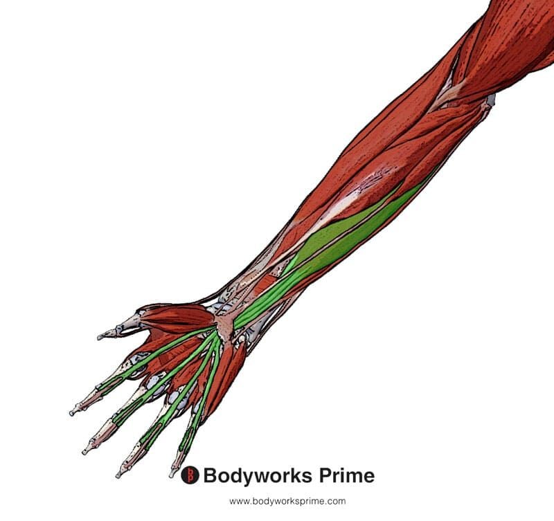
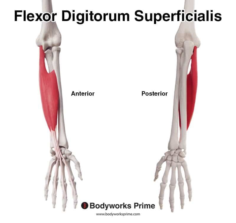
Flexor Digitorum Superficialis Muscle Anatomy Bodyworks Prime
Summary. origin: flexor digitorum superficialis tendon near the transverse carpal ligament insertion: metacarpal head of the index finger near the A1 pulley Gross anatomy. The accessory flexor digitorum superficialis muscle of the index finger is characterized by several variations that have been reported in the literature.
Flexor Digitorum Superficialis Muscle Anatomy Bodyworks Prime
Function. Flexor digitorum profundus muscle is a powerful flexor of the fingers. As it pulls the distal phalanges towards the hand, it causes flexion of the digits 2-5 at the metacarpophalangeal and interphalangeal joints.The muscle can act on its own but it usually works in synergy with the flexor digitorum superficialis, lumbricals and flexor digiti minimi brevis muscles to perform this action.
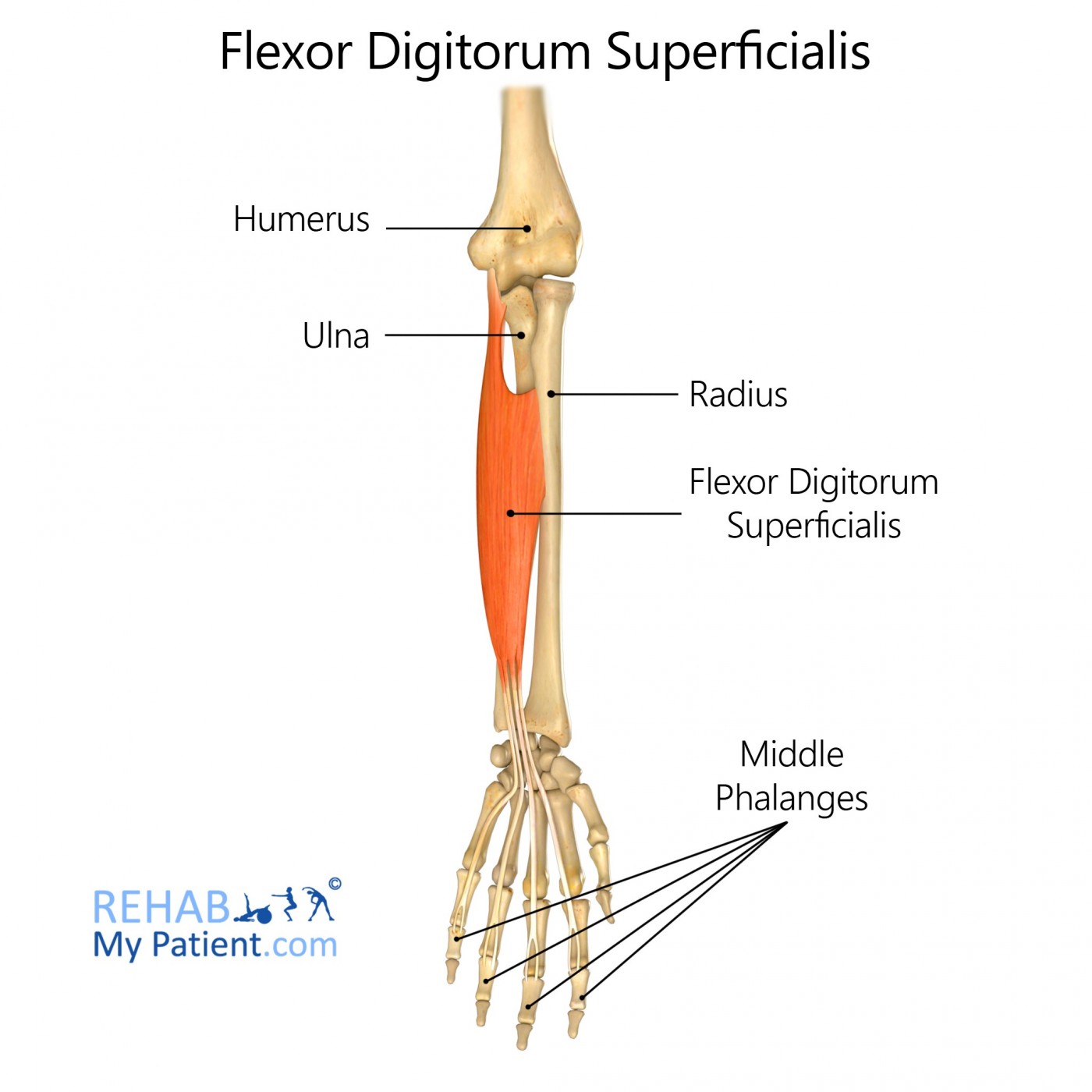
Flexor Digitorum Superficialis Rehab My Patient
The flexor digitorum superficialis muscle (FDS) is considered the most important of the forearm flexors for maintaining elbow valgus stability. However, the relationships between the origin structure of each finger of the FDS and the anterior oblique ligament (AOL) of the ulnar collateral ligament and the common tendon (CT) in the proximal.

Anatomy of Flexor Digitorum Superficialis —
Mori also found flexor digitorum superficialis joined with pronator teres 2.5% of subjects, flexor pollicis longus in 45%, and with flexor digitorum profundus in 8% of subjects (205 arms). Macalister reported the variations in flexor digitorum sublimis (superficialis) as follows: Absence of the tendon for the little finger by Wood and Macalister;
